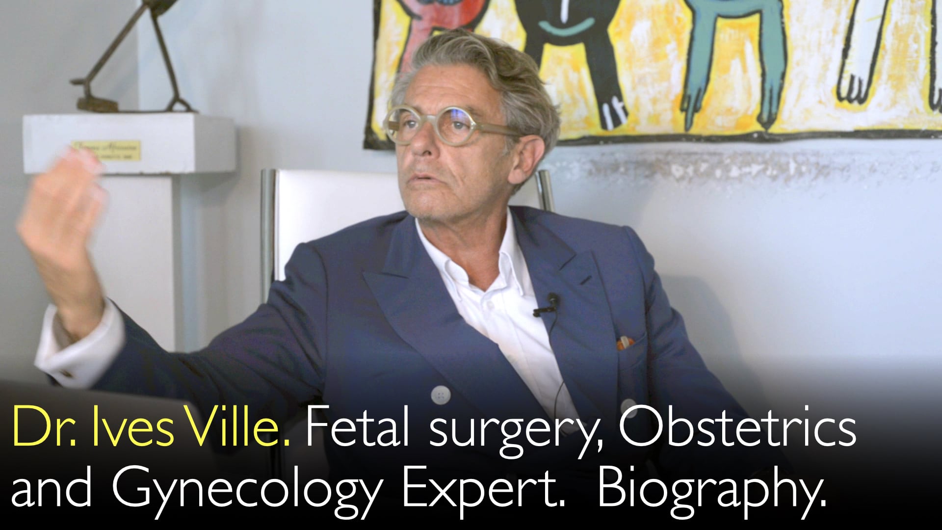
Leading expert in maternal-fetal medicine and endoscopic fetal surgery, Dr. Yves Ville, MD, explains how fetal interventions treat life-threatening congenital conditions. He details the evolution of minimally invasive techniques that operate on the placenta or fetus. These procedures dramatically improve survival for twins with twin-twin transfusion syndrome. Fetal surgery also treats severe congenital diaphragmatic hernia and rare, drug-resistant fetal arrhythmias. Dr. Yves Ville, MD, discusses the careful selection of conditions where the benefits outweigh the risks of preterm labor.
Endoscopic Fetal Surgery: Treating Congenital Abnormalities Before Birth
Jump To Section
- Twin-Twin Transfusion Syndrome Treatment
- Monochorionic Pregnancy Complications
- Congenital Diaphragmatic Hernia Repair
- Fetal Arrhythmia Intervention
- Spina Bifida Controversy
- Future Fetal Surgery Indications
- Full Transcript
Twin-Twin Transfusion Syndrome Treatment
Endoscopic fetal surgery first succeeded in treating twin-twin transfusion syndrome (TTTS). Dr. Yves Ville, MD, explains this condition affects monochorionic twins who share a single placenta. Abnormal vascular connections on the placental surface cause an imbalance of blood flow between the twins. The pioneering procedure involves coagulating these shared vessels using a micro-endoscope.
Dr. Yves Ville, MD, notes the pregnancy itself aids this surgery. The ample amniotic fluid provides a clear view of the operating field. This technique, developed in 1991, has become the global standard of care. It transformed outcomes from a 90% mortality rate to a 90% survival rate for affected twins.
Monochorionic Pregnancy Complications
Fetal surgery addresses several life-threatening complications in monochorionic pregnancies. Dr. Yves Ville, MD, describes selective intrauterine growth restriction (sIUGR). In this condition, one twin is severely growth-restricted and sick. Laser surgery can disconnect the circulations to save the healthier twin.
Another indication is selective termination for a lethal fetal anomaly. Surgeons can occlude the umbilical cord of the affected twin using laser or bipolar forceps. This spares the healthy co-twin. Dr. Ville also highlights TRAP sequence (Twin Reversed Arterial Perfusion). This involves coagulating the cord of a non-viable acardiac twin to prevent cardiac overload in the pump twin.
Congenital Diaphragmatic Hernia Repair
Severe congenital diaphragmatic hernia (CDH) is another key indication for fetal intervention. Dr. Yves Ville, MD, explains the pathophysiology. Abdominal organs herniate into the thorax through a diaphragmatic hole. This compresses the lungs and severely impairs their development.
The endoscopic procedure, called fetal endoscopic tracheal occlusion (FETO), temporarily blocks the fetal trachea. A balloon is placed inside the trachea to trap lung fluid. This increases intratracheal pressure and promotes lung growth. After three to four weeks, the balloon is removed. Dr. Anton Titov, MD, discusses the significant impact. This intervention can increase survival from 10% to 20-30% in the most severe cases.
Fetal Arrhythmia Intervention
Fetal surgery can treat rare, drug-resistant cases of fetal tachyarrhythmia. Dr. Yves Ville, MD, specifies this is for atrial flutter. This arrhythmia can lead to overt cardiac failure if pharmacological therapy fails. The endoscopic technique adapts tools used for other conditions.
Surgeons pass a small pacing probe into the fetus's esophagus behind the heart. They then pace the heart to stop the abnormal rhythm. Upon restarting, the heart often resumes a normal sinus rhythm. Dr. Yves Ville, MD, emphasizes these indications are extremely rare but demonstrate the versatility of the fetal surgical toolbox.
Spina Bifida Controversy
Fetal surgery for spina bifida represents a controversial expansion of indications. Dr. Yves Ville, MD, notes the fundamental principle of fetal surgery was to prevent death or severe irreversible sequelae. Spina bifida does not typically cause fetal death. The goal is to improve neurological outcomes, particularly motor function, but it does not cure incontinence.
The procedure involves endoscopically closing the myelomeningocele defect in utero. Dr. Ville observes a cultural and legal divide. In Europe, where termination is more accessible, this surgery is rare. In the US and other regions, it is offered more frequently as an alternative. This pushes the boundary of fetal surgery's core ethical framework.
Future Fetal Surgery Indications
The future of fetal surgery may include new conditions but faces significant obstacles. Dr. Yves Ville, MD, discusses sacrococcygeal teratoma, a highly vascular tumor. It can lead to fetal cardiac failure, but endoscopic attempts to treat it have so far failed. This may remain an indication for open fetal surgery, which involves opening the uterus.
Dr. Ville expresses a strong caution against open fetal surgery techniques. The maternal risks are substantial, including uterine rupture and placenta accreta in future pregnancies. He states a preference to develop only minimally invasive, endoscopic indications. This prioritizes maternal safety while advancing fetal care, a sentiment he believes is shared by many patients and practitioners.
Full Transcript
Dr. Anton Titov, MD: You are one of the leading experts in maternal-fetal medicine and fetal congenital abnormalities. You are also an expert on laser fetal surgery during pregnancy. What are typical medical problems that you solve for pregnant women? What diagnoses can be treated with endoscopic fetal surgery?
Dr. Yves Ville, MD: Right. So, the conditions have to be recognizable before treatment. There is a limited number of them. Chronologically, the first one used to actually try and then standardize fetal endoscopy was a condition that affects monochorionic twins. It's called twin-to-twin transfusion syndrome.
You're not operating on the fetuses themselves, but you're operating on the placenta. Twins share the same placenta. This placenta shows typical features of sharing part of the placenta through vessels running across the chorionic plates. Once you can recognize the pattern, you can also coagulate those vessels that are shared between the two fetuses. We did that in 1991 using micro-endoscopy that was developed for other purposes, for example, nose and throat surgery, but also for pediatric urology.
Those endoscopes are less than two millimeters in diameter. We found it acceptable to introduce that through a trocar percutaneously through the maternal abdomen into the uterus and into the amniotic fluid. The condition of pregnancy itself helps the surgery very much because it produces a lot of amniotic fluid, which gives you a very clear view of your operating field. That is not something that happens very often in the other conditions I'm going to discuss. But this one was particularly favorable.
Over the last nearly 30 years, this endoscopic fetal surgery technique has become the standard of care for this condition and other conditions related to monochorionicity. It means that two fetuses share the same placenta. Placental surgery was the first attempt and the first success.
That also pertains to selective growth restriction in one monochorionic twin. When one of those two twins, the small one, is very sick, it is sometimes life-saving, at least for the other twin, to disconnect the two twins. It is also sometimes necessary or acceptable to coagulate the umbilical cord of one twin when there is a lethal malformation in one twin.
So it's a selective termination of pregnancy by occluding the umbilical cord. This can be done either by laser or cord coagulation with bipolar forceps, but this is then under ultrasound guidance. So it's a vast spectrum of disease related to monochorionicity, where either you try to save both fetuses, like in twin-twin transfusion, or you want to make sure that the normal twin makes it through alive and well.
You have to then either separate from a very sick fetus or coagulate the cord of the very sick fetus. Or to coagulate a cord of the not quite a fetus, like the acardiac twin. The TRAP sequence twin, reversed arterial perfusion sequence, where there is a mass of fetal tissue, but it's neither an embryo nor a fetus. That is attached to a cord and is causing cardiac overload in an otherwise normal fetus next to it. So you have to disconnect this kind of tumor from the twin.
That's the whole thing about monochorionicity. In the world otherwise, this monochorionic disease spectrum represents probably 80% of fetal or intrauterine surgery.
Then you have other indications that were developed later on. One is related to diaphragmatic hernia, a congenital diaphragmatic hernia, where a hole in the diaphragm leaves the abdominal organs coming up from the abdomen into the thorax. This compresses the lungs, and the problem is lung development here.
To help struggle against the mechanical pressure of the viscera up in the thorax and allow more space for the lungs to grow, you can obstruct temporarily the trachea of the fetus. For this, it's fetal intubation. You use the same equipment as in twin-twin transfusion syndrome. You get into the uterus, the amniotic fluid, you open the mouth, you get into the trachea. You push a balloon that you blow up.
You leave it for three to four weeks to give time for those lungs to grow and benefit from the secretion of the terminal bronchial and the alveolar growth factors. Then you go back the same way, and you remove the balloon or the plug, as it's called. When this baby is delivered, you have optimized the chances for these babies to be able to breathe or be resuscitated.
I'd say in the most severe cases where survival is about 10%, it is now assumed that you increase survival two- or three-fold. So it's not a definite cure, but it's a great improvement. For twin-twin transfusion, we moved from 90% death to 90% survival, so the benefit is much clearer.
After the congenital diaphragmatic hernia, we thought we could use the same device in another condition, which is fetal arrhythmia, tachyarrhythmia. In a particular form of arrhythmia, that is, flutter. Flutter is a wrong connection between the auricles and the ventricles, where auricles beat much quicker than the ventricles. The connection between atria and ventricles happens only one in four or something like this.
Then that evolves into cardiac failure. When there is an overt cardiac failure, and the pharmacology therapy has failed—we have several drugs we can try to stop this—then we do use the same endoscopic instruments. Instead of targeting the trachea, we go into the esophagus. With the endoscope, we also pass a small probe, a pacing probe, the same pacing probe that pediatricians are using in these conditions after birth.
We go behind the heart behind the auricle that is beating wrongly, and we just pace the heart. The heart stops and then starts again. Usually, it starts again at the right rhythm. So we did that a couple of times. The indications are extremely rare. It's a rare problem. This problem is usually solved with medications, but for the resistant forms, we can use fetal surgery.
We have developed quite a toolbox based upon endoscopic access to the fetus. Then a team in Houston has used fetal endoscopy to close spina bifida in utero. This is a much more controversial indication because it's not a matter of life and death. What founded fetal surgery is a lethal condition if you don't treat before birth, or severe irreversible sequelae if you don't treat before birth.
Everything else really should be left to after birth because the disadvantage and the risk of fetal surgery are obviously by intruding the uterus; you can cause preterm labor. The very fundamental of fetal surgery was to prevent fetuses from dying. When we move to spina bifida, this is breaking that law because spina bifida will not be cured. The sequelae of spina bifida are still going to be present.
But a big clinical study showed that they might have some improvement in their motor capacity. Fetal surgery doesn't solve the incontinence problems in spina bifida. So several teams have moved on. In Europe, spina bifida indication is extremely rare, although spina bifida is not rare because people in Europe usually would terminate the pregnancy.
In the US and other parts of the world, where termination of pregnancy is not that available and less requested, this is an alternative that is being developed. We have a few cases that we do every year also here. We do that the same way with the endoscope in the uterus. But to facilitate the movements because now you're operating really on the features, we open the maternal abdomen but not the uterus.
We close the spina bifida with another port and put some stitches on it after dissecting the lesion. So those myelomeningoceles are just at the frontier of what fetal surgery is indicated for. There will probably be another few indications in the future for fetal endoscopy.
But one of the big failures or obstacles is a condition called a sacrococcygeal teratoma, a massive tumor of the sacrum and the coccyx. As sacrococcygeal teratoma name says, it's very vascular. It progresses very quickly towards cardiac failure. But such a big mass of tissue and so many vessels that so far, every single attempt by endoscopy or under ultrasound have failed.
That might be one of the last indications for intrauterine open surgery, where you open the uterus. Now, when you do that, again, you're entering another territory, which is the morbidity you're using in the woman. At least in our practice, that is something we don't want to cross. That's the line we don't want to cross.
We don't want to expose those women to a significant risk during this pregnancy or for the next pregnancy. There have been reports of this open fetal surgery. The sequelae of the uterus, the risk of uterine rupture, the risk of placenta accreta, the placenta that gets trapped into the scar. So we don't want to cross this line.
We are ready to develop any other indication on those could be, but we don't want to open the uterus. French women are not that keen on that either. Mostly. Probably it's cultural. And probably it's also legal, where in France, termination of pregnancy is allowed up to term when the fetal disease is severe.


![Endoscopic fetal surgery. When and where surgery on fetus can help? 1. [Parts 1 and 2]](http://diagnosticdetectives.com/cdn/shop/products/Dr_Ives_Ville_Obstetrics_fetal_surgery_pregnancy_infections_CMV_second_opinion_dr_anton_titov_Diagnostic_Detectives_Network_DiagnosticDetectives.Com.002.jpg?v=1660998401&width=1080)
![Endoscopic fetal surgery. When and where surgery on fetus can help? 1. [Parts 1 and 2]](http://diagnosticdetectives.com/cdn/shop/products/Dr_Ives_Ville_Obstetrics_fetal_surgery_pregnancy_infections_CMV_second_opinion_dr_anton_titov_Diagnostic_Detectives_Network_DiagnosticDetectives.Com.002_7d1d5dd7-18cb-46f8-84a8-7a549816b15d.jpg?v=1660998410&width=1080)
![Endoscopic fetal surgery. When and where surgery on fetus can help? 1. [Parts 1 and 2]](http://diagnosticdetectives.com/cdn/shop/products/Dr_Ives_Ville_Obstetrics_fetal_surgery_pregnancy_infections_CMV_second_opinion_dr_anton_titov_Diagnostic_Detectives_Network_DiagnosticDetectives.Com.002.jpg?v=1660998401&width=720)
![Endoscopic fetal surgery. When and where surgery on fetus can help? 1. [Parts 1 and 2]](http://diagnosticdetectives.com/cdn/shop/products/Dr_Ives_Ville_Obstetrics_fetal_surgery_pregnancy_infections_CMV_second_opinion_dr_anton_titov_Diagnostic_Detectives_Network_DiagnosticDetectives.Com.002_7d1d5dd7-18cb-46f8-84a8-7a549816b15d.jpg?v=1660998410&width=720)


