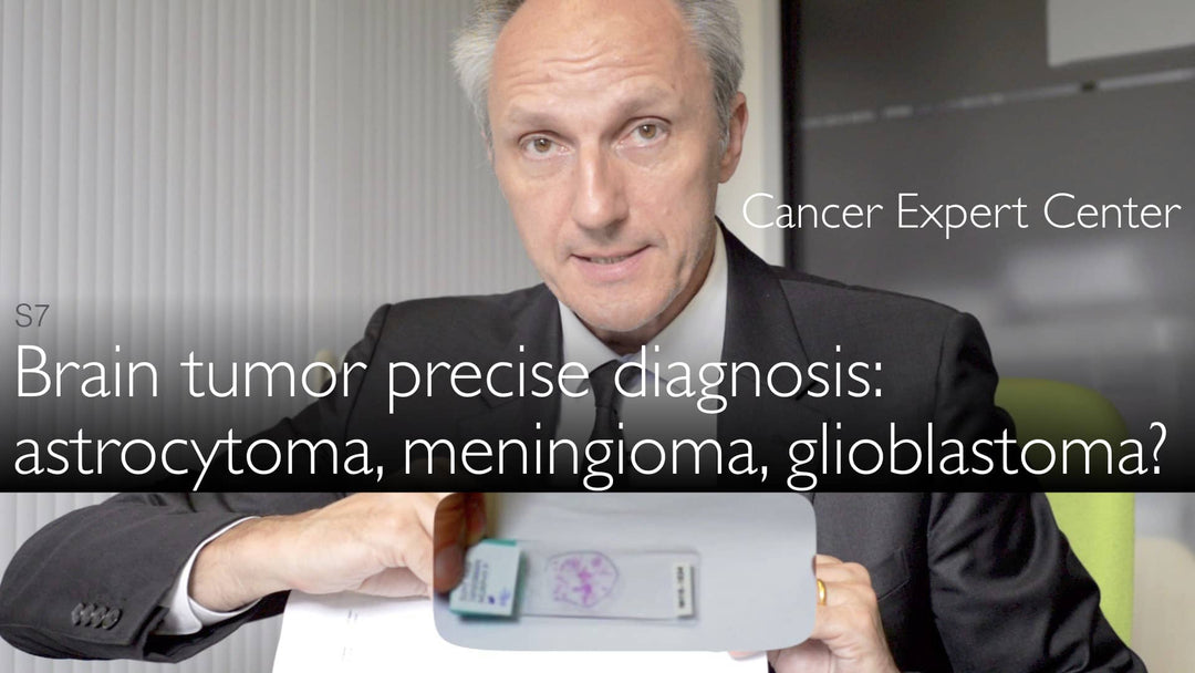
Leading expert in neuropathology, Dr. Sebastian Brandner, MD, explains how molecular diagnostics revolutionize brain tumor diagnosis and treatment. He details the critical role of IDH mutation testing in gliomas and astrocytomas while highlighting when traditional histology remains sufficient for meningioma diagnosis.
Molecular Diagnostics in Brain Tumor Pathology: From Meningioma to Glioblastoma
Jump To Section
- Traditional Histology in Brain Tumor Diagnosis
- Simple Diagnosis of Benign Meningiomas
- Molecular Testing for Gliomas and IDH Mutations
- Key Biomarkers in Astrocytoma Diagnosis
- Prognostic Factors Beyond Tumor Type
- The Future of Brain Tumor Diagnostics
- Full Transcript
Traditional Histology in Brain Tumor Diagnosis
Dr. Sebastian Brandner, MD, emphasizes that basic histology remains the foundation of brain tumor pathology. The initial H&E (hematoxylin and eosin) stained slide provides crucial first insights, with pink-stained tissue samples revealing tumor architecture under microscopy. This traditional method continues to guide the diagnostic pathway for many brain tumor cases.
Simple Diagnosis of Benign Meningiomas
Meningiomas arising from brain coverings often require no molecular testing, as Dr. Sebastian Brandner, MD, explains. These typically benign tumors can be definitively diagnosed through standard histology alone. The neuropathologist notes that tumor location - particularly difficult-to-access skull base positions - often impacts prognosis more than molecular factors in meningiomas. Surface meningiomas generally have excellent outcomes with complete surgical resection.
Molecular Testing for Gliomas and IDH Mutations
For gliomas, especially in younger patients, Dr. Sebastian Brandner, MD, highlights the critical importance of IDH mutation testing. A specialized antibody developed at the German Cancer Research Center detects 90% of IDH1 mutations and rare IDH2 variants. This cost-effective immunohistochemical method provides rapid, reliable results that significantly influence treatment decisions and prognosis assessment.
Key Biomarkers in Astrocytoma Diagnosis
Dr. Brandner describes a two-antibody approach for astrocytoma diagnosis. The IDH mutation test combines with analysis of nuclear protein loss - when the characteristic "black dot" disappears from cell nuclei, it strongly indicates astrocytoma. This molecular signature helps differentiate astrocytomas from other glioma subtypes and guides appropriate treatment strategies.
Prognostic Factors Beyond Tumor Type
While molecular diagnostics provide essential information, Dr. Brandner stresses that tumor location and surgical accessibility remain crucial prognostic factors. Even benign tumors in challenging anatomical positions may have poorer outcomes than more aggressive tumors in operable locations. The neuropathologist emphasizes the need for comprehensive evaluation combining histological, molecular and clinical data.
The Future of Brain Tumor Diagnostics
Dr. Sebastian Brandner, MD, anticipates continued advancement in molecular pathology techniques. The success of IDH mutation testing demonstrates how targeted biomarkers can streamline diagnosis while improving accuracy. As research identifies more tumor-specific molecular signatures, pathology laboratories will increasingly combine traditional and molecular methods for optimal patient care.
Full Transcript
Dr. Anton Titov, MD: Previously, brain tumors were diagnosed relatively crudely. Basic staining is still the first step in the pathology analysis of brain cancer. But now there is more molecular diagnostics and molecular analysis of brain tumor mutations. This becomes very important for treatment of tumors. It is important for the brain tumor prognosis.
Dr. Anton Titov, MD: What is the importance of molecular diagnostics for brain tumor diagnosis and therapy?
Dr. Sebastian Brandner, MD: First of all, you are absolutely correct that the mainstay of pathology diagnostics is still the first histology slide. It is a slide that looks like this. It has pink staining. I just give an example. This is the size of a slide - 1 by 3 inches. Sometimes you can see against the background of these little pink flics in the center. These are the brain tumor tissue specimen.
Dr. Sebastian Brandner, MD: We put this slide first under the microscope. That is the first decision in brain tumor diagnosis.
Dr. Anton Titov, MD: What to do next?
Dr. Sebastian Brandner, MD: First of all, there might be a number of tumors, like the benign meningioma. Sometimes a brain tumor arises from the coverings of the brain rather than from within the brain. Meningiomas are usually diagnosed just on this section slide. So it's a fast and cheap brain tumor diagnosis. It gives the best information.
Dr. Sebastian Brandner, MD: There is no need to do any further molecular testing. Because there are other factors determining whether this brain tumor will have bad prognosis or a good prognosis. For example, where the brain tumor grows. Sometimes it grows in difficult to reach areas. Bottom of the brain, which is called "skull base". Then this determines the clinical prognosis.
Dr. Sebastian Brandner, MD: The brain tumor that is easily accessible on the surface of the brain can be taken out. Usually these brain tumors don't recur. If meningiomas recur, they can often be resected a second time.
Dr. Sebastian Brandner, MD: These are the meningiomas. You can diagnose them just on single H&E slide. Gliomas generally can also be diagnosed on a single H&E simple staining slide, particularly glioblastomas.
Dr. Sebastian Brandner, MD: But there are patients who are younger. Some patients have gliomas that have mutations that I mentioned earlier. Gliomas have IDH mutation. These IDH mutations can be detected with an antibody. This antibody was generated in the laboratory in Heidelberg at the German Cancer Research Center. This antibody detects the glioma mutation. So it's a cheap and quick, pathologist-friendly way to diagnose mutations in a brain tumor.
Dr. Sebastian Brandner, MD: The nice thing is that this antibody detects 90% of all IDH mutations. 95% of IDH1 mutations and very rare, so-called IDH2 mutations in glioma. This antibody is then combined with another antibody. That antibody detects a nuclear protein. It should normally be present in the cell in the nucleus.
Dr. Sebastian Brandner, MD: But in some type of brain tumors this protein is lost in the nucleus. These brain tumors are astrocytomas. So the small black dot is lost from the cell's nucleus. We would normally find this black dot in the middle of the cell, in the nucleus. That loss is characteristic and nearly diagnostic for astrocytoma.
Dr. Anton Titov, MD: IDH mutation is present also in astrocytoma brain tumor. Brain tumor diagnosis expert explains precise molecular diagnosis of brain tumor type. Benign meningioma. More aggressive astrocytoma and glioblastoma multiforme (GBM).


